Dental radiographs (X-rays) are an instrumental diagnostic tool in modern dentistry, they offer not only valuable information that is invisible during a regular dental exam but also provide a way for practitioners to detect disease problems and corruption far earlier than if left ignored. The dental practice in Glen Oaks, NY uses many different kinds of X-rays to help spot dental issues and monitor treatment plans. In this article, we will discuss the types of dental X-rays available for use and how they are done on a patient.
Dental X-Rays
Dental X-rays provide visibility into areas that are not visible to the naked eye, including cavities, infections, bone loss, and tumors. They are also important to plan for treatments such as fillings, crowns, braces, and dental implants. Routine X-rays provide an important snapshot of your oral health and can help monitor changes in dental health over time.
Types of Dental X-Rays
1. Bitewing X-Rays
It is used to show the upper and lower posterior teeth of one area in your mouth like as top left molar, it helps see cavities where they most commonly form. These are the X-rays that show decay between teeth and changes in bone density, such as those caused by gum disease. They are invariably taken during regular dental exams and can show the condition of dental restorations. the patient bites down on a film or digital sensor that takes images of the teeth in this location.
2. Periapical X-Rays
This X-ray concentrates on a particular tooth and shows the entire tooth, its root, and surrounding bone. These X-rays are used to diagnose issues such as abscesses, cysts, or infections at the root of a tooth, as well as monitor the health of bone around the tooth. The CR sensor or film is inserted into the mouth and an X-ray image is captured of one or two teeth at a time.
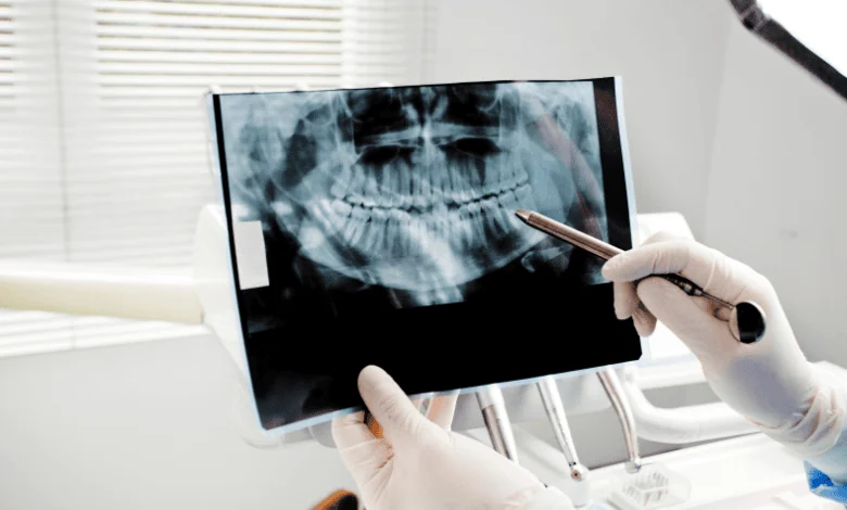
3. Panoramic X-Rays
It reveals an extensive picture of the complete mouth, including all of the teeth, jaws, and surrounding structures. The purpose of these pictures is to help plan orthodontic treatments, diagnose impacted teeth, and evaluate general oral health. They can also aid in the detection of jaw problems or malignancies. The patient is positioned to either stand or sit within a machine that revolves around their head, taking a single picture that shows all of their teeth and facial features at once.
4. Cone Beam Computed Tomography
CBCT is a form of X-ray that provides detailed, 3D views of the teeth, soft tissues, and bone. The 3D imaging and diagnosed areas X-ray are mainly for complex cases, such as implant planning, the evaluation of jaw structure, or the extent of tumors in malignant disease. The client rests in a rotating maker that takes numerous images that combine to create a three-dimensional design.
5. Occlusal X-Rays
These images show the top view of the teeth in the lower or upper jaw. Usually, they are employed to evaluate tooth alignment and identify anomalies in the growth of the jaws and teeth, such as cysts or additional teeth. To take an image of the occlusal surface of the teeth, the film or sensor is inserted into the mouth and the patient is allowed to bite down on it.
By learning more about what dental X-rays are and the available different types, patients in Glen Oaks, NY can understand why this diagnostic tool is so important for oral health. Dental X-rays help detect problems early before symptoms appear, and they can provide valuable information about the health of teeth and their supporting structures which are essential for both treatment planning and preventive care. If you have any questions about dental X-rays, or your dental health in general be sure to speak with your dentist who will be able to give advice and care based on you.
Want more tips? Check out our blog.